Stochastic biophysical modeling and simulations (eg membrane dynamics)
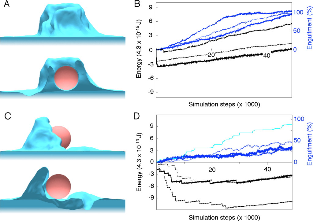
Simulations of a computational model coupling cell membrane thermodynamics (blue) with active biochemical processes to support cell membrane fluctuations. The model was designed to describe quantitatively how phagocytes (immune cells) engulf particles (e.g. pathogenic bacteria), using the zipper mechanism. (A) Side view (top) and cross section (bottom) of a phagocytic cup obtained for the active zipper. (B) Corresponding time courses of the membrane energy (thick solid, medium dashed and thin dotted black lines) and percentage of engulfment, defined by the average membrane height around the particle (blue solid, blue dashed, blue dotted lines) for three repeats of the stochastic simulation. (C) Side view (top) and cross section (bottom) of the phagocytic cup obtained for the passive zipper (same overall s imulation time). (D) Corresponding time courses of the membrane energy (thick solid, medium dashed and thin dotted black lines) and engulfment (blue solid, blue dashed, blue dotted lines) for three repeats of the simulation. Dashed light blue line indicates corresponding maximal membrane height for one of the simulations (in percentage of the particle diameter). Small particles with 1.5 μm radius were used. From Tollis et al BMC 2010
Biomathematical analysis of membrane receptors dynamics and signal to noise ratio optimization
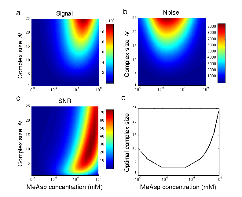
Optimal receptor complex size in the presence of both extrinsic ligand and intrinsic methylation noise (signaling noise). (a) Signal, (b) Noise, and (c) signal to noise ratio (SNR) as a function of ligand concentration and complex size of Tar receptors. (d) There is a maximum of the SNR at complex sizes larger than one receptor. Parameter values are given in appendix B. From Aquino et al, PRE 2011
Dynamical modeling of biochemical pathways (deterministic modeling)
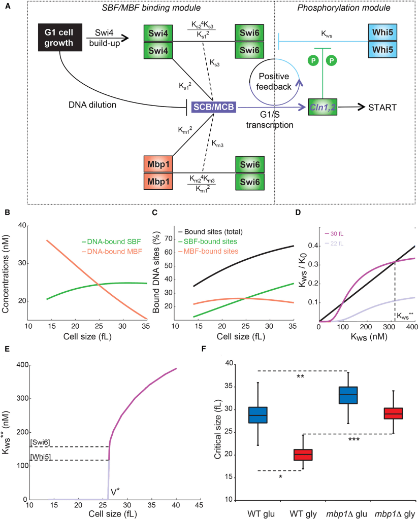
A Mathematical Model of the commitment to cell division in yeast. (A) Schematic of the most relevant biomolecular interactions. In the SBF/MBF binding module, the concentrations of Mbp1, Swi6, and Whi5 are size independent. Swi4 and Mbp1 can bind target promoter DNA either free of Swi6 (solid lines) or as fully formed hetero-tetramer SBF or MBF complexes (dotted lines).The output from the SBF/MBF binding module (i.e., concentrations of DNA-bound and DNA-free SBF/MBF) is used as an input in the phosphorylation module, which is solved for the Whi5-SBF dissociation constant, Kws. An increase in the fraction of uninhibited SBF-bound DNA target sites increases the likelihood of CLN1/2 transcription and thus amplification of phosphorylation on Whi5 and Swi6, which increases Kws and thereby releases more Whi5 from SBF at G1/S promoters to trigger a switch-like transition to cell division. (B) The concentrations of SBF bound by DNA (green) and MBF bound by DNA (orange) versus cell size. (C) The fraction of total G1/S promoter sites bound by SBF and MBF (black), MBF alone (orange) or SBF alone (green) versus cell size. (D) Graphical illustration of bistability in this model, where the solutions of the model are the intersections of black and purple curves. As cells grow from small (light purple) to large (dark purple) sizes, solutions appear in the high phosphorylation-large Kws region of the diagram, corresponding to activation of cell division. (E) Kws as a function of cell size. Whi5-SBF complex becomes unstable at a critical size V* where SBF activity and cell division are triggered. (F) Box and whisker plots of simulated critical cell size for WT and mbp1D cells in glucose (blue) versus glycerol (red) for nuclear concentrations of Swi4, Swi6, Mbp1, and Whi5 randomly picked within the ranges constrained by experimental measurements of absolute concentrations of the transcription factors (see also Quantitative Bio-Imaging). From Dorsey, Tollis et al Cell Systems 2018
Dynamical modeling at single molecule level (stochastic simulations)
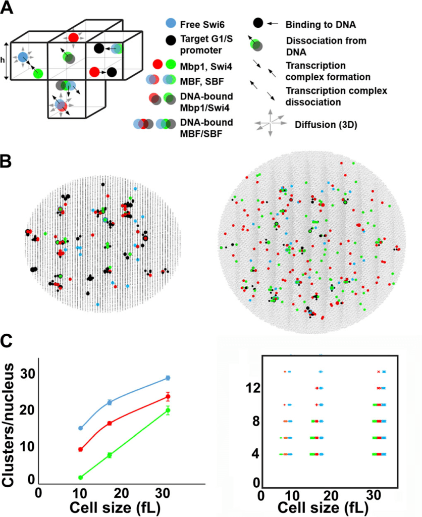
Stochastic 5space-dependent Gillespie) modeling predicts clustering of the Swi4, Mbp1, and Swi6 G1/S transcription factor molecules in budding yeast. (A) Schematic of the model (left), encompassing mobile Swi4 dimers (green dots), Mbp1 dimers (red dots), Swi6 dimers (blue dots), and immobile G1/S DNA promoters (black dots),moving and interacting in the nucleus discretized in infinitesimal volume elements. For illustrative purposes, the leftmost element shows Swi4 and Swi6 binding (convergent thick black arrows). The bottommost element shows dissociation of Mbp1 from Swi6 (divergent thick black arrows) and diffusion (thin gray arrows). Also shown is Swi4-Swi6 dissociation from DNA. The rightmost element shows Mbp1 and Swi4-Swi6 association with two promoters within a cluster. Propensities of single biochemical reactions can be calculated. 2D projection of the 3D output of a typical simulation showing clusters of Swi4 dimers (green dots), Mbp1 dimers (red), Swi6 dimers (blue), and G1/S DNA promoters (black dots) in small (10 fl, left) and large (31.5 fl, right) cells. (C) Left: mean number of Swi4 (green), Mbp1 (red), and Swi6 (cyan) clusters per nucleus (vertical axis) as a function of the size of 10 simulated cells (horizontal axis). Right: Scatter plot showing the number of Swi4 (green),Mbp1 (red), and Swi6 (cyan)molecules per cluster (vertical axis) as a function of the size of 10 simulated cells (horizontal axis). From Black, Tollis et al JCB 2020
Dynamical modeling of coupled multi-scale biochemical (molecules) and biophysical (vesicles) systems
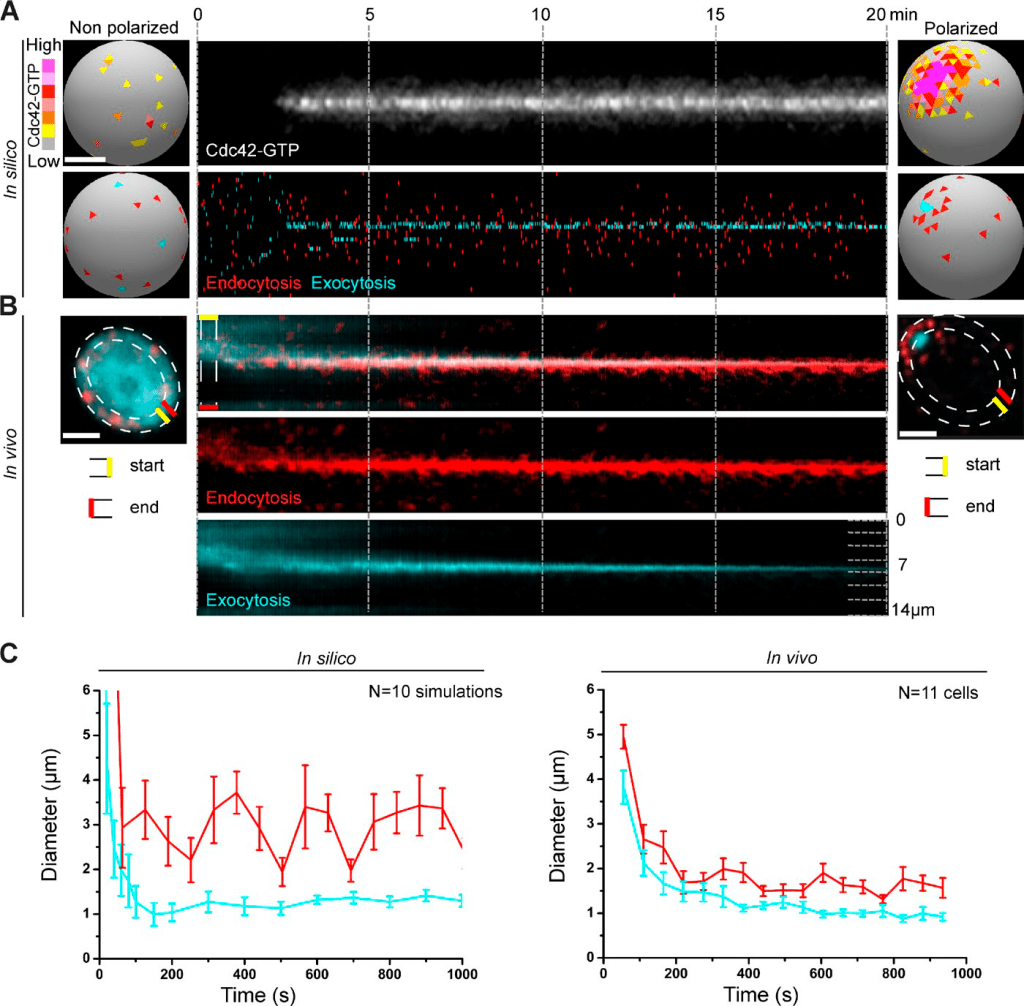
Robust cell polarization involves dynamic changes in endo- and exocytic vesicular trafficking systems. Predictions of a stochastic model of self-organization of endo and exocytic vesicles, alongside other membrane-bound cell polarity factors (constituting the Cdc42 GTPase module). (A) Transition of a typical in silico WT cell from nonpolarized to a polarized state. Membrane-bound Cdc42-GTP depolarized over the plasma membrane (top left) polarizes to a unique cluster over time (top right). The Cdc42 kymograph (top) shows Cdc42-GTP during polarization. The kymograph in the bottom shows individual endo and exocytic events over time (x axis) along the cortex (y axis). A tight pole of exocytosis develops (cyan), overlapping the Cdc42-GTP cluster, and is corralled by a ring of endocytosis (red). (B) Random endo- and exocytic distributions observed in vivo in a nonpolarized cell (left) change to an organized “bull’s-eye pattern” in a polarized cell (right) with a tight exocytic zone surrounded by endocytic vesicles, as predicted by the model. The endo- and exocytic zones are marked by Abp1-RFP (red) and GFP-Sec4 (cyan), respectively. Kymographs represent the endo- and exocytic profiles along the cortex. Bud emergence occurs at the end of the kymographs. Bars, 2 μm. (C) In silico and in vivo statistical analyses of the coincident tightening of the exocytic (cyan) and endocytic (red) zones as WT cells polarize with the SD between cells represented by error bars. From Jose, Tollis et al JCB 2013
Mathematical modeling of the effects of drug combinations against Acute Myeloid Leukemia (AML)
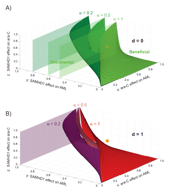
Mathematical analysis identifies quantitative conditions for a beneficial effect of specifically targeting SAMHD1’s ara-CTPase function. A) State diagram of the AML/ara-C/SAMHD1/anti-SAMHD1 drug system for d→0 (poorly effective drug), delineating regions of the (x,y,z) space where targeting SAMHD1 is beneficial for a poorly specific drug (α=1, light green), a moderately specific drug (α=0.5, medium green), and a highly specific drug (α=0,2, dark green). For each value of ɑ , the (x,y,z) region below/above the corresponding limit surface corresponds to situations where targeting SAMHD1 is detrimental/beneficial, respectively. B) State diagram of the AML/ara-C/SAMHD1/anti-SAMHD1 drug system for d=1 (very effective drug), delineating regions of the (x,y,z) space where targeting SAMHD1 is beneficial for a poorly specific drug (α =1, red), a moderately specific drug (α=0.5, dark pink), and a highly specific drug (α=0,2, violet). For each value of ɑ, the (x,y,z) region below/above the corresponding limit surface corresponds to situations where targeting SAMHD1 is detrimental/beneficial, respectively. Work in peer-review.